急性缺血性卒中OCTA表现与功能结局相关性数据
该数据集来自tandf.figshare.com,配套论文发表于Neuro-Ophthalmology(2024年),探讨光学相干断层扫描血管成像(OCTA)表现能否预测颅外颈动脉粥样硬化性疾病(ECAD)相关卒中的功能结局
基本信息
资源简介
急性缺血性卒中OCTA表现与功能结局相关性数据
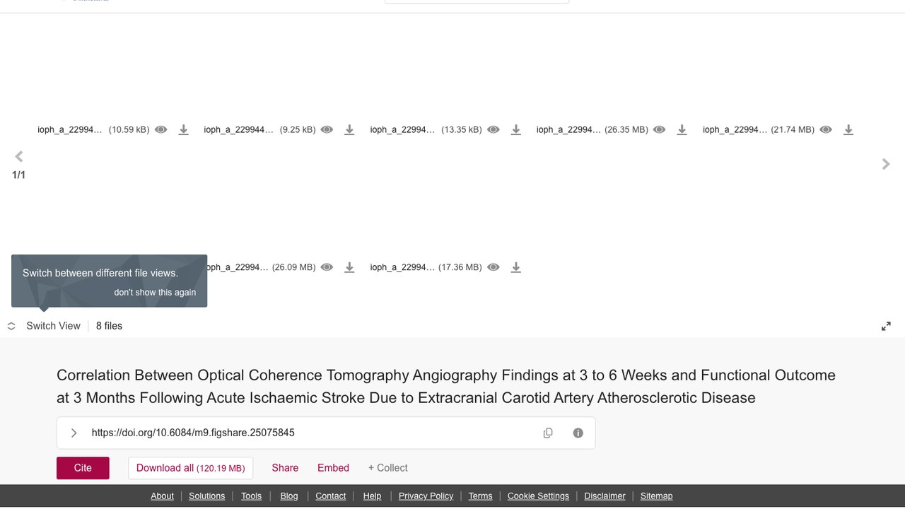
Correlation Between Optical Coherence Tomography Angiography Findings at 3 to 6 Weeks and Functional Outcome at 3 Months Following Acute Ischaemic Stroke Due to Extracranial Carotid Artery Atherosclerotic Disease
该数据集来自tandf.figshare.com,配套论文发表于Neuro-Ophthalmology(2024年),探讨光学相干断层扫描血管成像(OCTA)表现能否预测颅外颈动脉粥样硬化性疾病(ECAD)相关卒中的功能结局。研究纳入23例急性缺血性卒中患者与23例危险因素匹配的无颈动脉狭窄对照,于卒中后3~6周采集OCTA数据,包含8个补充文件(docx与tiff格式)。数据可用于分析黄斑深层血管密度、视盘周围放射状毛细血管灌注密度及浅层血管密度与3个月功能结局的关联,支持卒中预后预测研究(来源 figshare,CC BY 4.0 许可)。
【数据集背景】
配套论文《Correlation Between Optical Coherence Tomography Angiography Findings at 3 to 6 Weeks and Functional Outcome at 3 Months Following Acute Ischaemic Stroke Due to Extracranial Carotid Artery Atherosclerotic Disease》发表于 Neuro-Ophthalmology(2024),DOI:10.1080/01658107.2023.2299442。项目资助方:The author(s) reported there is no funding associated with the work featured in this article.。源站首次发布:2024-01-26。
【数据内容】
源站描述(机器翻译,仅供参考):我们希望评估光学相干断层扫描血管成像(OCTA)表现能否预测颅外颈动脉粥样硬化性疾病(ECAD)相关卒中的功能结局。这项探索性研究在因ECAD导致急性缺血性卒中的成人中进行,于卒中发病后3~6周采集数据,并设置危险因素匹配的无颈动脉狭窄对照。共纳入23例卒中患者(病例)和23例对照。病例与对照在黄斑深层血管密度(p = .0007)和视神经乳头(ONH)处放射状视乳头周围毛细血管灌注密度(RPCPD)(p = .0007)方面存在显著差异。在同侧眼黄斑总浅层血管密度(SVD)(SVD在对照数据1个标准差[SD]以内 versus SVD超出1个SD)与功能结局之间观察到统计学显著差异。
【数据结构与技术规格】
文件数 8 个,合计 126.03 MB。数据集 DOI:10.6084/m9.figshare.25075845.v1。
【主题与分类】
源站学科分类:Physical Sciences not elsewhere classified、Medicine、Cell Biology、Genetics、Neuroscience、Physiology。主题标签:Optical coherence tomography angiography、acute ischaemic stroke、modified Rankin scale。本站分类:循环系统疾病。
【适用方向】
源站学科分类标注为 Physical Sciences not elsewhere classified、Medicine、Cell Biology、Genetics、Neuroscience、Physiology。
【获取与许可】
源站页面:https://tandf.figshare.com/articles/dataset/Correlation_Between_Optical_Coherence_Tomography_Angiography_Findings_at_3_to_6_Weeks_and_Functional_Outcome_at_3_Months_Following_Acute_Ischaemic_Stroke_Due_to_Extracranial_Carotid_Artery_Atherosclerotic_Disease/25075845
使用许可:CC BY 4.0(https://creativecommons.org/licenses/by/4.0/)。
规范引用:Correlation Between Optical Coherence Tomography Angiography Findings at 3 to 6 Weeks and Functional Outcome at 3 Months Following Acute Ischaemic Stroke Due to Extracranial Carotid Artery Atherosclerotic Disease. Taylor & Francis. Mishra, Biswamohan; Pandit, Awadh Kishor; Chawla, Rohan; Aalok, Swat…
【使用注意】
· 源站最后修改于 2024-06-24,引用时请注意版本时效。
下载信息
注册下载
需要注册 Kaggle 账号并登录后下载,适合需要跟踪下载记录和使用 API 的用户。
暂未开放公开下载
无需注册即可直接获取公开样本或文档,适合快速预览和评估数据集质量。
免登录有偿下载
公开数据集受托下载与技术交付服务。
提供高速下载与技术交付服务(收技术服务费,非数据销售)
暂未开放当前数据集主要来源为 Kaggle 公开托管,完整影像包建议通过原始链接或 Kaggle API 获取。
使用方式
数据集获取(Figshare)
- 源站标题:Correlation Between Optical Coherence Tomography Angiography Findings at 3 to 6 Weeks and Functional Outcome at 3 Months Following Acute Ischaemic Stroke Due to Extracranial Carotid Artery Atherosclerotic Disease
- DOI:10.6084/m9.figshare.25075845.v1
- 发布日期:2024-01-26
- 文件数:8 个
- 文件总体积:126.03 MB
- 源站页面:https://tandf.figshare.com/articles/dataset/Correlation_Between_Optical_Coherence_Tomography_Angiography_Findings_at_3_to_6_Weeks_and_Functional_Outcome_at_3_Months_Following_Acute_Ischaemic_Stroke_Due_to_Extracranial_Carotid_Artery_Atherosclerotic_Disease/25075845
命令行下载
curl -L -o "ioph_a_2299442_sm6249.docx" "https://ndownloader.figshare.com/files/44244108"
curl -L -o "ioph_a_2299442_sm6242.docx" "https://ndownloader.figshare.com/files/44244111"
curl -L -o "ioph_a_2299442_sm6240.docx" "https://ndownloader.figshare.com/files/44244114"
curl -L -o "ioph_a_2299442_sm6216.tiff" "https://ndownloader.figshare.com/files/44244117"
curl -L -o "ioph_a_2299442_sm6201.tiff" "https://ndownloader.figshare.com/files/44244120"
curl -L -o "ioph_a_2299442_sm6154.tiff" "https://ndownloader.figshare.com/files/44244123"
curl -L -o "ioph_a_2299442_sm5553.tiff" "https://ndownloader.figshare.com/files/44244126"
curl -L -o "ioph_a_2299442_sm5288.tiff" "https://ndownloader.figshare.com/files/44244129"
文件清单
ioph_a_2299442_sm6249.docx(10.85 KB)ioph_a_2299442_sm6242.docx(9.47 KB)ioph_a_2299442_sm6240.docx(13.67 KB)ioph_a_2299442_sm6216.tiff(27.63 MB)ioph_a_2299442_sm6201.tiff(22.80 MB)ioph_a_2299442_sm6154.tiff(30.01 MB)ioph_a_2299442_sm5553.tiff(27.36 MB)ioph_a_2299442_sm5288.tiff(18.21 MB)
文件总体积 126.03 MB(源站 API 实测)。
数据加载示例(图像类)
from PIL import Image
import glob, os

files = (glob.glob(os.path.join(path, "**", "*.png"), recursive=True)
       + glob.glob(os.path.join(path, "**", "*.jpg"), recursive=True)
       + glob.glob(os.path.join(path, "**", "*.tif"), recursive=True))
print("图像文件数:", len(files))
img = Image.open(files[0]); print("尺寸/模式:", img.size, img.mode)

# torchvision Dataset 方式:
# from torchvision import datasets
# ds = datasets.ImageFolder(path)  # 要求 子目录=类别
目录组织与标注格式以源站说明和下载后实际文件为准。
数据说明
我们希望评估光学相干断层扫描血管成像(OCTA)检查结果能否预测颅外颈动脉粥样硬化性疾病(ECAD)相关卒中的功能结局。这项探索性研究在因ECAD导致急性缺血性卒中的成人中进行,于卒中发病后3–6周纳入,并设置危险因素匹配的无颈动脉狭窄对照组。共纳入23例卒中患者(病例组)和23名对照者。病例组与对照组在黄斑深部血管密度(p = .0007)及视神经乳头(ONH)处径向视乳头周围毛细血管灌注密度(RPCPD)(p = .0007)方面存在显著差异。在同侧眼黄斑总浅层血管密度(SVD)(SVD在对照数据1个标准差[SD]以内 versus SVD超出1个SD)与3个月时功能结局(结局不良 versus 结局极好,分别为改良Rankin量表[mRS] 0–1分 versus mRS 2–6分)之间观察到统计学显著差异(p = .0361)。
视盘处RPCPD与入院时美国国立卫生研究院卒中量表评分、出院时mRS及卒中发病后3个月时mRS之间均无统计学显著相关性(分别为r=0.33,r=0.35,r=0.39;p=0.11,p=0.09,p=0.06)。这项探索性研究的结果提示,OCTA检查结果或可预测ECAD相关卒中的3个月结局,并可能有助于未来干预研究在决定是否对存在关键性或非关键性ECAD的患者进行干预以预防卒中时作出决策。
许可
CC BY 4.0
引用
学术使用请引用 DOI 10.6084/m9.figshare.25075845.v1(source: https://tandf.figshare.com/articles/dataset/Correlation_Between_Optical_Coherence_Tomography_Angiography_Findings_at_3_to_6_Weeks_and_Functional_Outcome_at_3_Months_Following_Acute_Ischaemic_Stroke_Due_to_Extracranial_Carotid_Artery_Atherosclerotic_Disease/25075845)。
数据缺失?
依托陕西、四川两大基地,我们与超过十家三甲医院建立直接合作关系,覆盖合规授权、采集治理、专业标注、数据交付的全流程,为AI医疗团队提供即拿即用的高质量临床数据。
⚡️ 需要数据支持或标注服务?立即联系我们获取专业方案。